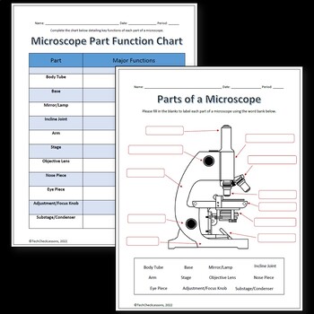
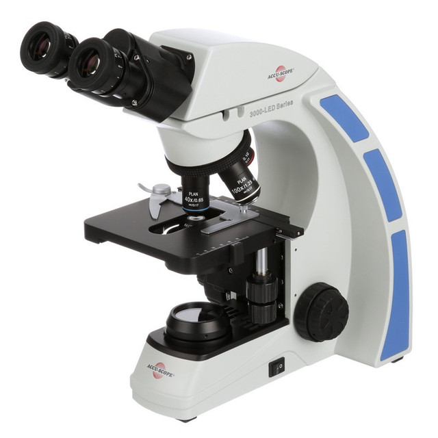
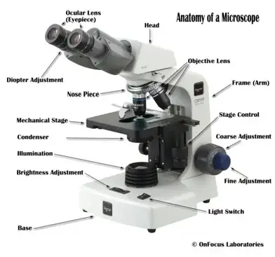
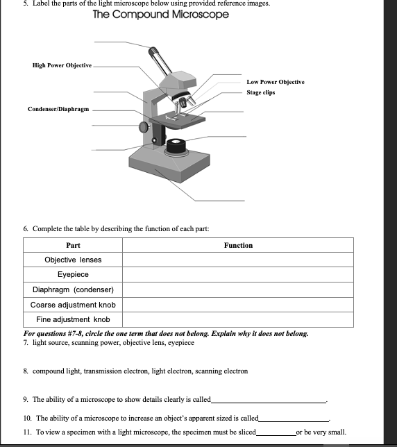
39 label the image of a compound light microscope using the terms provided
BIO 168 Module 2 Quiz Review Flashcards | Quizlet Drag each label to the appropriate layer (A, B, or C) for each term or phrase. ... Label the image of a compound light microscope using the terms provided. ... Use fine focus to sharpen the image under low power. 5. Rotate high power lens into position. 6. Readjust fine focus under high power to produce the sharpest image. Bio 232 ~ Lab Midterm Flashcards - Quizlet Match each cellular component to whether it is bound by a membrane or not by dragging each label into the appropriate box. Not all terms will be used. Label the anatomy of the mitochondrion in the figure below. Label the image of a compound light microscope using the terms provided. ... Label the skeletal system components in the figure with ...
Microscopy Lab Quiz Flashcards | Quizlet Label the image of a compound light microscope using the terms provided. If leaving an objective lens over the stage when storing the microscope, which objective lens should be placed over the stage? A. High power 40X
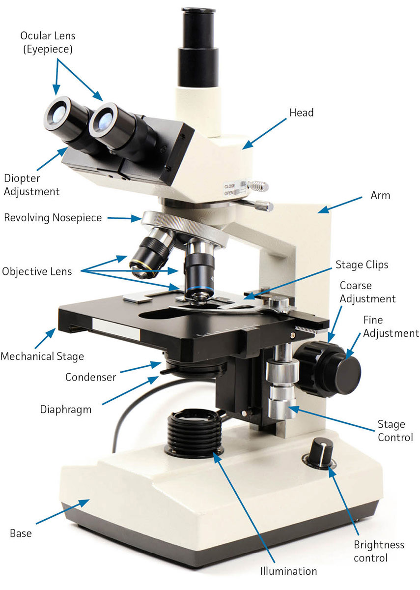
Label the image of a compound light microscope using the terms provided
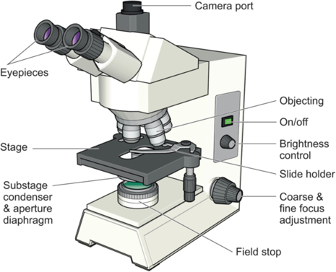
Compound Microscope Parts - Labeled Diagram and their Functions - Rs ... There are two major optical lens parts of a microscope: Eyepiece (10x) and Objective lenses (4x, 10x, 40x, 100x). Total magnification power is calculated by multiplying the magnification of the eyepiece and objective lens. The illuminator provides a source of light. The light is focused by the condenser and passing through the specimen placed ... compound microscope parts (labeling) Flashcards | Quizlet Start studying compound microscope parts (labeling). Learn vocabulary, terms, and more with flashcards, games, and other study tools. ... light source of the microscope. what is 8? eyepiece (ocular lens) - magnifying piece that is looked into in order to see the specimen ... knob that brings the image to a sharper focus. what is 13? base - the ... Online Lab 2 The Compound Light Microscope.docx - Name:... View Online Lab 2 The Compound Light Microscope.docx from BIOL 216 at Eastern University. Name: Lizbeth Ramos Microbiology The Compound Light Microscope Please label the diagram using the terms below. Study Resources. Main Menu ... Name: Lizbeth Ramos Microbiology The Compound Light Microscope Please label the diagram using the. Online Lab 2 ...
Label the image of a compound light microscope using the terms provided. Solved Label the image of a compound light microscope using - Chegg Expert Answer. Labeling of the Left Side blanks of microscope are as follows A. Rotating Nosepiece B. Objective Lenses C. Slide Holder Finger D. Stage E. Condenser F. Iris Diaphragm lever G. Substage illuminator (lamp) H. Mechanical Stage Control knob Labeling of t …. View the full answer. Transcribed image text: Label the image of a compound ... Label the above components of the compound light microscope A Ocular ... Keiser University Online Microbiology I Virtual Lab 1 Worksheet Microscopy & Gram Staining microscope b. resolution The power to show details clearly. The optic ability to distinguish detail such as the separation of closely adjacent objects. c. contrast converts phase shifts in light passing through a transparent specimen to brightness changes in the image Virtual Lab 2 Microscopy and Cells_for Hybrid absense.docx... Virtual Lab 2 - Microscopy and Cells BIO101 General Biology I ACTIVITY 1: MICROSCOPY Read the Introduction on page 23 of the lab manual. The Dissecting Microscope (aka: stereoscope): Watch the following YouTube video on the parts, function, and usage of the Leica EZ4 dissecting microscope (which is the model we have in the BIO101 lab rooms): o Page 23, bottom: Label the following image of a ... Parts of a microscope with functions and labeled diagram Q. Differentiate between a condenser and an Abbe condenser. Ans. Condensers are lenses that are used to collect and focus light from the illuminator into the specimen. They are found under the stage next to the diaphragm of the microscope. They play a major role in ensuring clear sharp images are produced with a high magnification of 400X and above.
Label the image of a compound light microscope using the terms provided. 1. State a procedures which should be used to properly handle a light microscope. 2. Explain why the light microscope is also called the compound microscope. 3- Images observed under the light microscope are reversed and inverted. Explain what this... Compound Microscope - Diagram (Parts labelled), Principle and Uses Also called as binocular microscope or compound light microscope, it is a remarkable magnification tool that employs a combination of lenses to magnify the image of a sample that is not visible to the naked eye. Compound microscopes find most use in cases where the magnification required is of the higher order (40 - 1000x). Compound Microscope: Definition, Diagram, Parts, Uses, Working ... - BYJUS A compound microscope is defined as. A microscope with a high resolution and uses two sets of lenses providing a 2-dimensional image of the sample. The term compound refers to the usage of more than one lens in the microscope. Also, the compound microscope is one of the types of optical microscopes. The other type of optical microscope is a ... Labelled Diagram of Compound Microscope - Biology Discussion The below mentioned article provides a labelled diagram of compound microscope. Part # 1. The Stand: The stand is made up of a heavy foot which carries a curved inclinable limb or arm bearing the body tube. The foot is generally horse shoe-shaped structure (Fig. 2) which rests on table top or any other surface on which the microscope in kept.
Compound Light Microscope Diagram Worksheet - Google Groups Study manual following chapter which describes features of the initial light microscope and the function of each carbon the diagram of the microscope below. You will label sketches to compound light microscope worksheet may want to your students to use worksheets to. On a typical student compound light microscope there are 3-4 of objective lenses. Solved Label the image of a compound light microscope using - Chegg Step-by-step answer. Who are the experts? Experts are tested by Chegg as specialists in their subject area. We review their content and use your feedback to keep the quality high. Transcribed image text: Label the image of a compound light microscope using the terms provided. PDF The Compound Light Microscope lab - Caldwell-West Caldwell Public Schools Part B: Care and handling of a compound light microscope Procedure: Answer the following questions concerning the care and handling of a compound light microscope. 1. Why is it important to carry the microscope correctly? 2. Why does the microscope need to be set at least 5cm away from the edge of the table? 3. Microscope parts/labeling Label the image of a compound light ... 1. State a procedures which should be used to properly handle a light microscope. 2. Explain why the light microscope is also called the compound microscope. 3- Images observed under the light microscope are reversed and inverted.
PDF The Compound Light Microscope Storing the Microscope Turn the objective lens to lowest power, lower the stage, & remove the slide Turn off the light, unplug the cord, & wrap the cord neatly around the microscope Use two hands to carry the microscope back to the storage area Place the protective bag over the microscope before you store it away Scientific Drawing Checklist
Label the image of a compound light microscope using the terms provided. Microscope parts/labeling 9 Label the image of a compound light microscope using the terms provided. 1 points eyepiece eyepiece light source References References base arm slide holder arm stage mechanical stage fine adjustment knob power switch...
A & P Microscope and Cell cycle Flashcards - Quizlet Study with Quizlet and memorize flashcards terms like What is the typical magnification provided by the eyepiece (ocular) lens system?, The greatest magnification on the compound light microscope can be achieved by using the high power objective lens., A parfocal microscope is one that keeps the specimen in focus (or very close to it) when a higher-power objective lens is rotated into position ...
PDF Label the image of a compound light microscope light microscope using the terms provided. Many important anatomic features, especially those that work on the levels of tissues or cell phones, are too small to be seen by the eye without help. The composite microscope is a valuable tool to expand small sections of biological material so that inaccessible details can be solved.
Labeling the Parts of the Microscope Labeling the Parts of the Microscope. This activity has been designed for use in homes and schools. Each microscope layout (both blank and the version with answers) are available as PDF downloads. You can view a more in-depth review of each part of the microscope here.
Parts of the Microscope with Labeling (also Free Printouts) Picture Source: magnet.fsu.edu. 8. Illuminator. It illuminates the light to the specimen for better visualization. It is the one responsible for creating light rays for magnification. Image 8: The encircled part of the microscope is the illuminator. Picture Source: alicdn.com. 9. Condenser.
Online Lab 2 The Compound Light Microscope.docx - Name:... View Online Lab 2 The Compound Light Microscope.docx from BIOL 216 at Eastern University. Name: Lizbeth Ramos Microbiology The Compound Light Microscope Please label the diagram using the terms below. Study Resources. Main Menu ... Name: Lizbeth Ramos Microbiology The Compound Light Microscope Please label the diagram using the. Online Lab 2 ...
compound microscope parts (labeling) Flashcards | Quizlet Start studying compound microscope parts (labeling). Learn vocabulary, terms, and more with flashcards, games, and other study tools. ... light source of the microscope. what is 8? eyepiece (ocular lens) - magnifying piece that is looked into in order to see the specimen ... knob that brings the image to a sharper focus. what is 13? base - the ...
Compound Microscope Parts - Labeled Diagram and their Functions - Rs ... There are two major optical lens parts of a microscope: Eyepiece (10x) and Objective lenses (4x, 10x, 40x, 100x). Total magnification power is calculated by multiplying the magnification of the eyepiece and objective lens. The illuminator provides a source of light. The light is focused by the condenser and passing through the specimen placed ...













Post a Comment for "39 label the image of a compound light microscope using the terms provided"