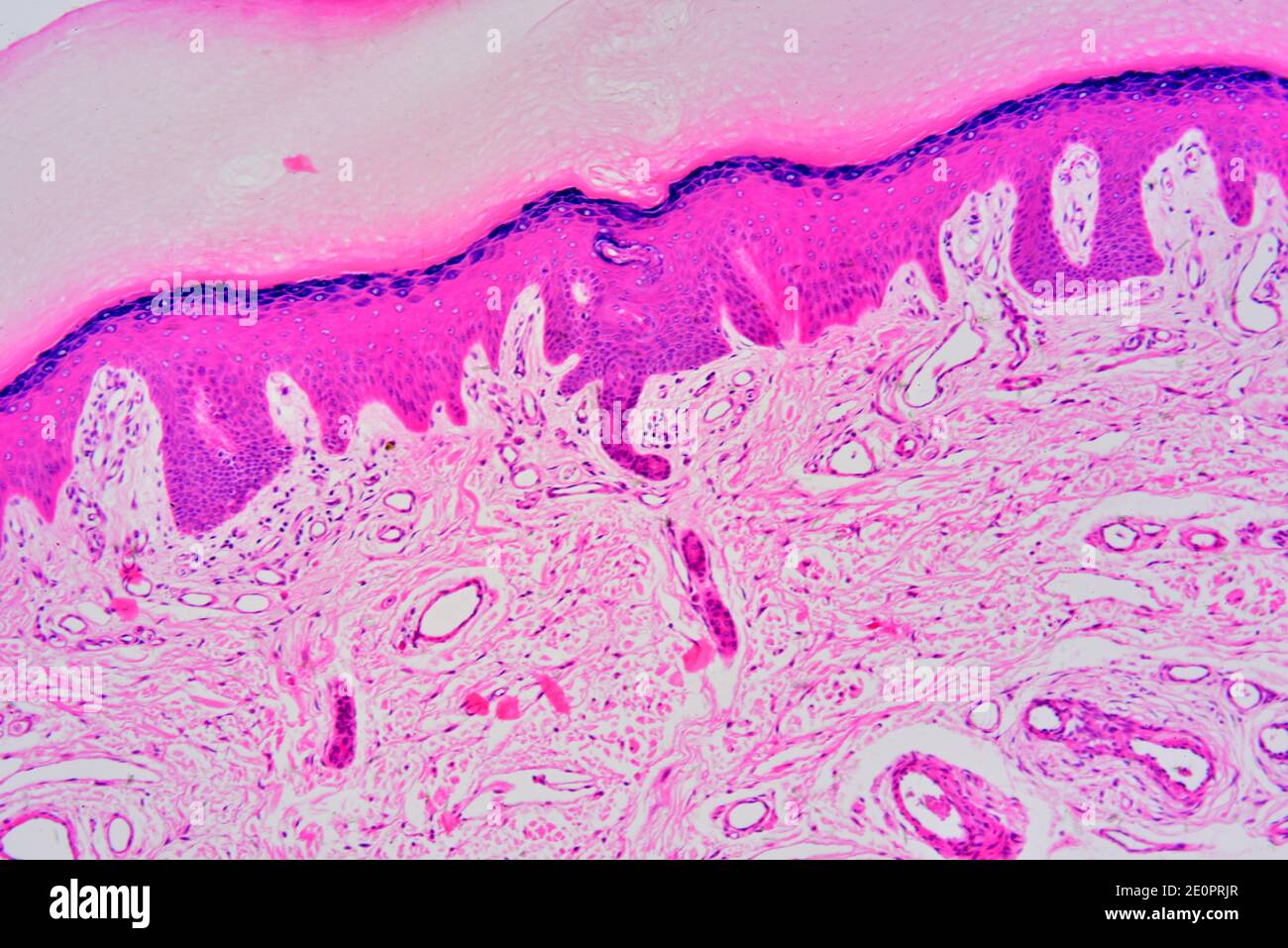
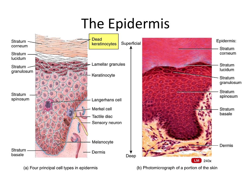
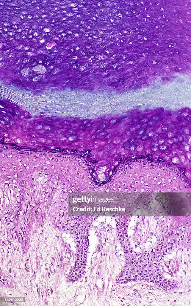
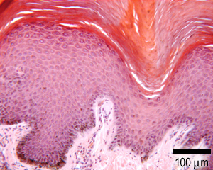
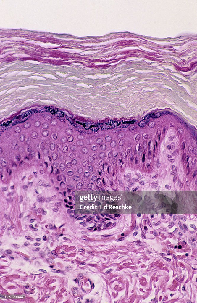
45 photomicrograph of thick skin
photomicrograph of thick skin Diagram - Quizlet Start studying photomicrograph of thick skin. Learn vocabulary, terms, and more with flashcards, games, and other study tools. Photomicrograph of Thick Skin Quiz - PurposeGames This online quiz is called Photomicrograph of Thick Skin. It was created by member nhammond21 and has 6 questions.
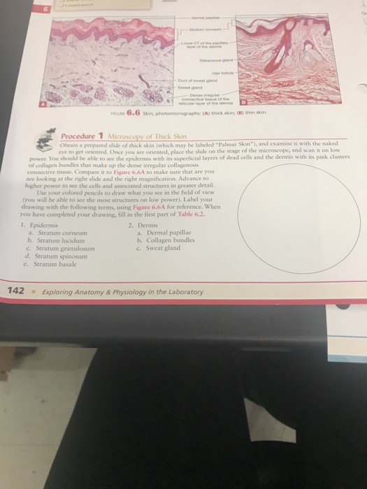
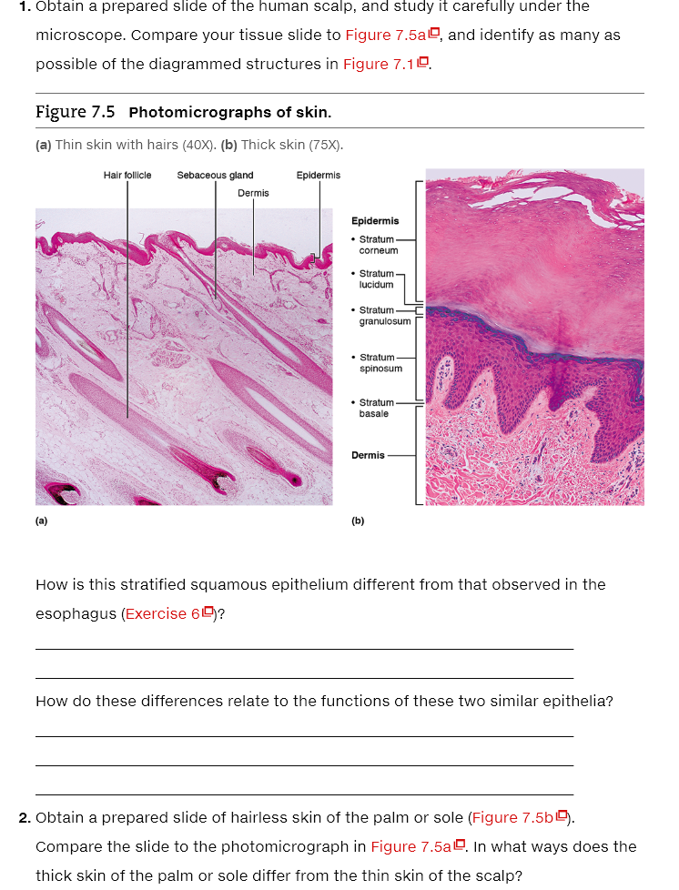
(a): A photomicrograph of the section of thin skin tissue from the... Note the hair follicles (HF), sebaceous glands (SG), and well-defined dense collagenous tissue (DCT) (H&E, 10x). (b) A photomicrograph of H&E section of thin ...

Photomicrograph of thick skin
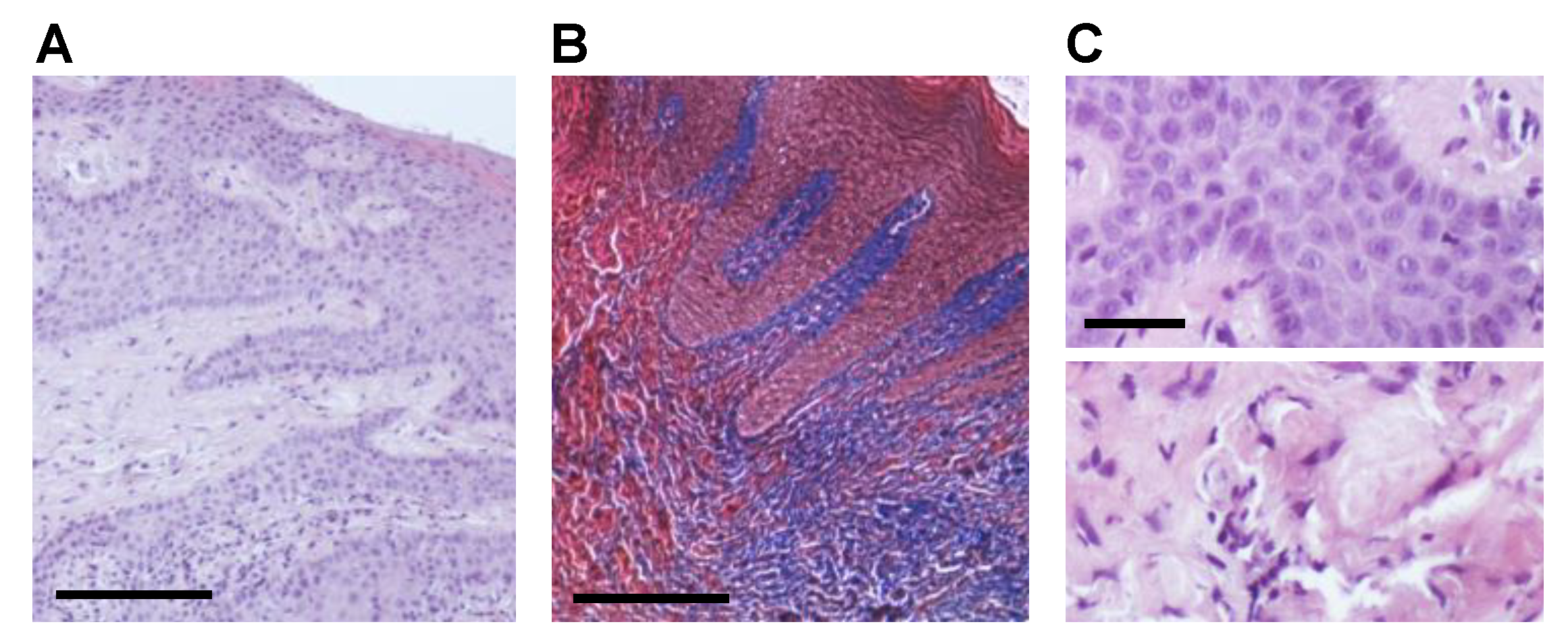
Photomicrographs of dermis from dorsal skin of the four-striped ... Surface grooves were evident in the cornified layer of the trunk skin, but not in the paw pad skin. The mean thickness of the cornified layer of the epidermis ... 5.1 Layers of the Skin – Anatomy & Physiology Part A is a micrograph showing a cross section of thin skin. The topmost layer Figure 5.1.2 – Thin Skin versus Thick Skin: These slides show cross-sections of ... Block1/Fig 10. Dermis of thick skin Fig 10. Dermis of thick skin. This photomicrograph shows the connective tissue of the skin, referred to as dermis, stained to show the nature and ...
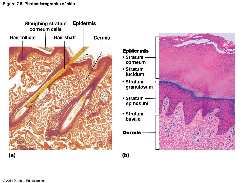
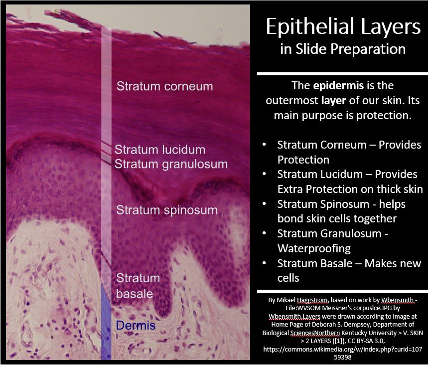
Photomicrograph of thick skin. Skin overview 4 | Digital Histology A diagrammatic representation of thin skin and a photomicrograph of a H&E stained section illustrate the reduced thickness of the epidermal strata in thin ... The differences between thick and thin skin - The Histology Guide Can you identify the five major layers of the epidermis? Dermis: Thick skin has a thinner dermis than thin skin, and does not contain hairs, sebaceous glands, ... Solved Label the photomicrograph of thick skin. Stratum | Chegg.com Question: Label the photomicrograph of thick skin. Stratum corneum Stratum basale Stratum granulosum Stratum lucidum Epidermis Dermis Stratum spinosum. Label the photomicrograph of thick skin. Stratum corneum Stratum ... The Label of the photomicrograph of thick skin is given in the image attached. What factors affect skin thickness?
Block1/Fig 10. Dermis of thick skin Fig 10. Dermis of thick skin. This photomicrograph shows the connective tissue of the skin, referred to as dermis, stained to show the nature and ... 5.1 Layers of the Skin – Anatomy & Physiology Part A is a micrograph showing a cross section of thin skin. The topmost layer Figure 5.1.2 – Thin Skin versus Thick Skin: These slides show cross-sections of ... Photomicrographs of dermis from dorsal skin of the four-striped ... Surface grooves were evident in the cornified layer of the trunk skin, but not in the paw pad skin. The mean thickness of the cornified layer of the epidermis ...














Post a Comment for "45 photomicrograph of thick skin"