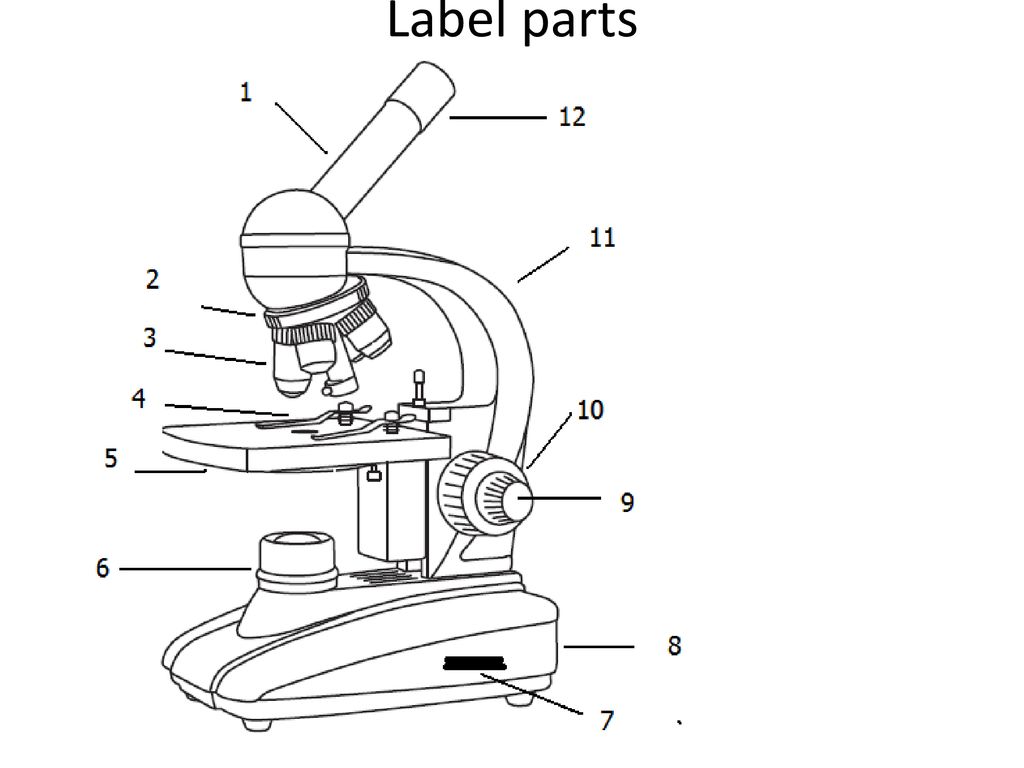
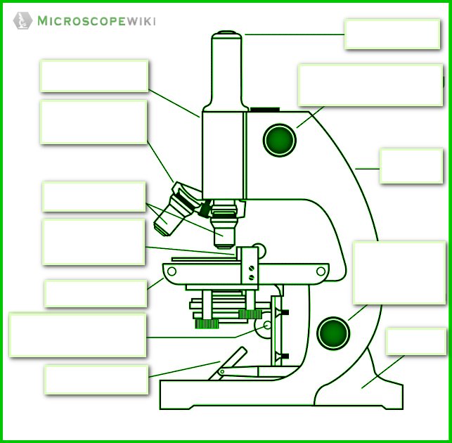
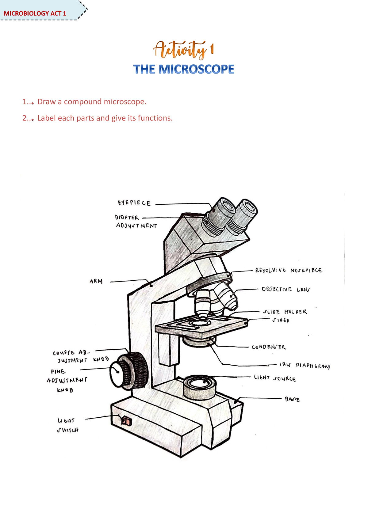
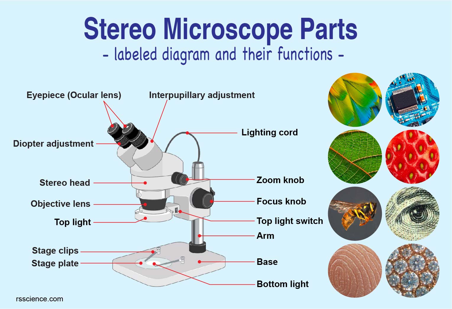
42 picture of compound microscope with label
3.1: Introduction to the Microscope - Biology LibreTexts A compound light microscope has a maximum resolution of 0.2 µm, this means it can distinguish between two points ≥ 0.2 µm, any objects closer than 0.2um will be seen as 1 object. ... This introduction to microscopy will include an explanation of features and adjustments of a compound brightfield light microscope, which magnifies images ... 19,831 Microscope Drawing Images, Stock Photos & Vectors - Shutterstock Find Microscope Drawing stock images in HD and millions of other royalty-free stock photos, illustrations and vectors in the Shutterstock collection. Thousands of new, high-quality pictures added every day.
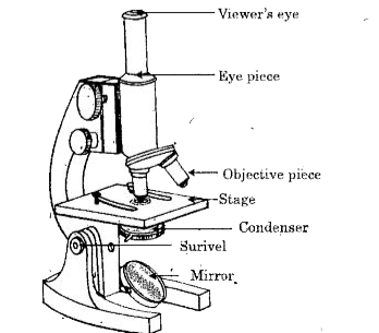
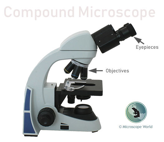
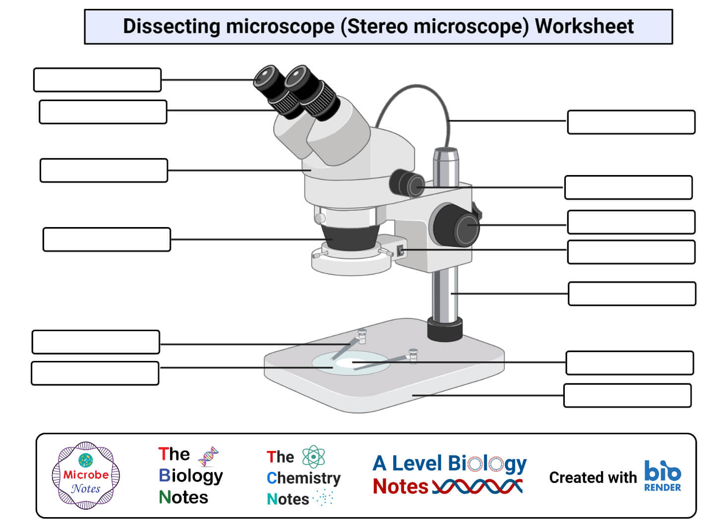
Parts of a microscope with functions and labeled diagram - Microbe Notes The optical parts of the microscope are used to view, magnify, and produce an image from a specimen placed on a slide. These parts include: Eyepiece - also known as the ocular. This is the part used to look through the microscope. Its found at the top of the microscope.

Picture of compound microscope with label
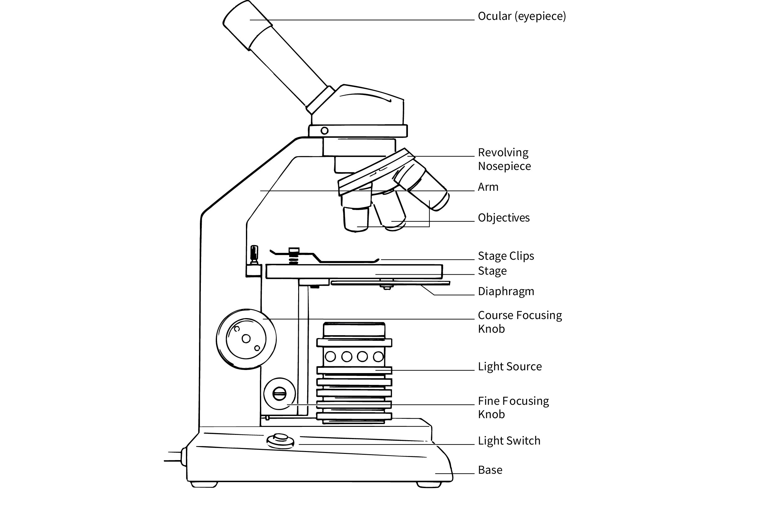
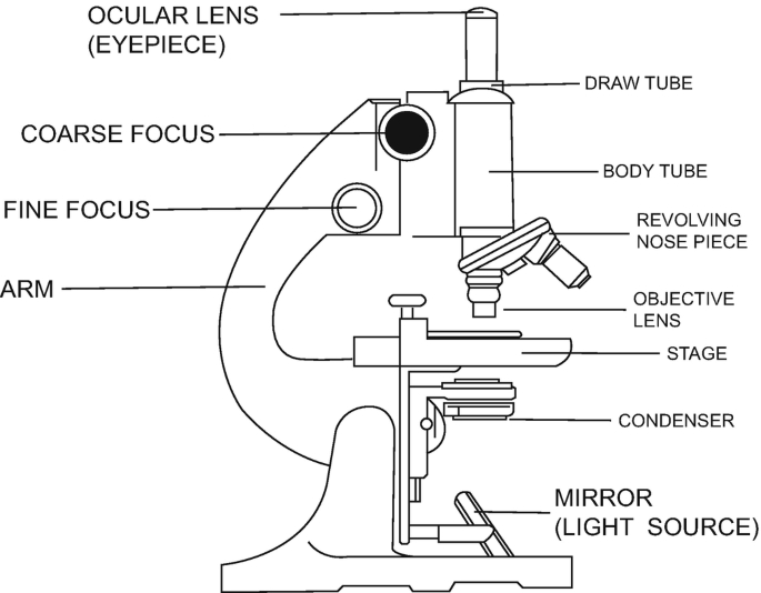
Compound Microscope Parts - Labeled Diagram and their Functions Labeled diagram of a compound microscope Major structural parts of a compound microscope There are three major structural parts of a compound microscope. The head includes the upper part of the microscope, which houses the most critical optical components, and the eyepiece tube of the microscope. Compound Microscope Labeled Diagram | Quizlet Compound Microscope Labeled + − Flashcards Learn Test Match Created by meganplocher734 Terms in this set (14) Eyepiece/Ocular lens Contains the ocular lens Body tube A hollow cylinder that holds the eyepiece. Arm Part that supports the microscope. Stage Supports the slide or specimen Coarse adjustment Knob Compound Microscope Photos and Premium High Res Pictures - Getty Images a view down the eye piece of a upright compound microscope - compound microscope stock pictures, royalty-free photos & images people-centric innovation in industrial research and development. clinical laboratory scientist is using a microscope in a laboratory of pharmacy industry. - compound microscope stock pictures, royalty-free photos & images
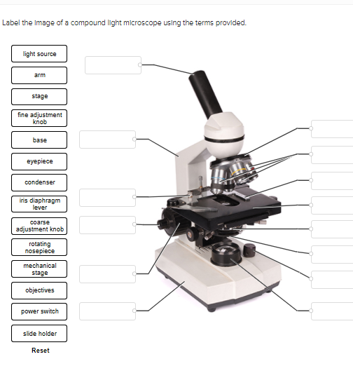
Picture of compound microscope with label. Compound Microscope - Diagram (Parts labelled), Principle and Uses A compound microscope basically consists of optical and structural components. Within these two systems, there are multiple components within them and they are: Image : Labeled Diagram of compound microscope parts See: Labeled Diagram showing differences between compound and simple microscope parts Structural Components Label the microscope — Science Learning Hub Label the microscope Interactive Add to collection Use this interactive to identify and label the main parts of a microscope. Drag and drop the text labels onto the microscope diagram. diaphragm or iris base eye piece lens fine focus adjustment light source stage coarse focus adjustment high-power objective Download Exercise Compound Microscope- Definition, Labeled Diagram, Principle, Parts, Uses Compound microscopes have a combination of lenses that enhances both magnifying powers as well as the resolving power. The specimen or object, to be examined is usually mounted on a transparent glass slide and positioned on the specimen stage between the condenser lens and objective lens. 1.5: Microscopy - Biology LibreTexts Gently scrape the inside of your cheek with a toothpick and swirl it in the dye on the slide. Place a cover slip on the suspension and view at 1000X total magnification. Draw 1-3 cells large enough to show the detail that you see in your lab manual. Label its cell membrane, cytoplasm and nucleus.
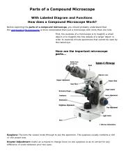
Parts of a Compound Microscope - Labeled (with diagrams) Image 1: The figure above is the standard image of a compound microscope. image source: 5.imimg.com The structural components of a compound microscope. Picture 2: The basic parts of a compound microscope. image source : optimaxonline.com Head/body it is where the upper optical parts of the microscope can be found. Base Compound Microscope: Definition, Diagram, Parts, Uses, Working Principle A compound microscope is defined as A microscope with a high resolution and uses two sets of lenses providing a 2-dimensional image of the sample. The term compound refers to the usage of more than one lens in the microscope. Also, the compound microscope is one of the types of optical microscopes. Microscope Parts and Functions Microscope Parts and Functions With Labeled Diagram and Functions How does a Compound Microscope Work?. Before exploring microscope parts and functions, you should probably understand that the compound light microscope is more complicated than just a microscope with more than one lens.. First, the purpose of a microscope is to magnify a small object or to magnify the fine details of a larger ... Microscope Types (with labeled diagrams) and Functions Compound microscope labeled diagram Compound microscope functions: It finds great application in areas of pathology, pedology, forensics etc Its greater order of magnification allows for deeper study of microbial organisms to Detect the cause of diseases Study the mineral composition in soils
Diagram of a Compound Microscope - Biology Discussion 1. It is noted first that which objective lens is in use on the microscope. 2. Stage micrometer is positioned in such a way that it is in the field of view. 3. The eyepiece is rotated so that the two scales, the eyepiece or ocular scale and the stage micrometer scale, are parallel. 4. Compound Microscope Labeled Definition, Labeled Diagram, Best procedure ... How to Work Compound Microscopes. A compound microscope or compound microscope labeled is a type of microscope that uses two or more lenses to magnify an object. The first lens, called the eyepiece, is used to view the object. The second lens, called the objective lens, is used to collect light from the object and focus it on the eyepiece. Microscopy: Intro to microscopes & how they work (article) - Khan Academy This picture isn't a plain light micrograph; it's a fluorescent image of a specially prepared plant where various parts of the cell were labeled with tags to make them glow. However, this kind of cellular complexity and beauty is all around us, whether we can see it or not. Compound Microscope Parts, Functions, and Labeled Diagram Compound Microscope Definitions for Labels Eyepiece (ocular lens) with or without Pointer: The part that is looked through at the top of the compound microscope. Eyepieces typically have a magnification between 5x & 30x. Monocular or Binocular Head: Structural support that holds & connects the eyepieces to the objective lenses.
Parts of the Microscope (Labeled Diagrams) - Simple and Compound Microscope Parts of Compound Microscope (Labeled Pictures) a. Mechanical Parts of a Compound Microscope Foot or Base Pillar Arm Stage Inclination Joint Clips Diaphragm Nose piece/Revolving Nosepiece/Turret Body Tube Adjustment Knobs b. Optical Parts of a Compound Microscope Eyepiece lens or Ocular Mirror Objective Lenses Scanning Objective Lens (4x)
Microscopes | Digital, Compound, Stereo | Fisher Scientific Compound microscopes are used to view images of small objects on glass slides. Magnification can range from 40x to 1000x and is achieved by multiple convex lenses in the eyepieces and in magnifying objectives. ... Digital microscopes create digital or electronic images and may be any type of microscope (compound, inverted, stereo, etc.). Before ...
Compound Microscope Photos and Premium High Res Pictures - Getty Images a view down the eye piece of a upright compound microscope - compound microscope stock pictures, royalty-free photos & images people-centric innovation in industrial research and development. clinical laboratory scientist is using a microscope in a laboratory of pharmacy industry. - compound microscope stock pictures, royalty-free photos & images
Compound Microscope Labeled Diagram | Quizlet Compound Microscope Labeled + − Flashcards Learn Test Match Created by meganplocher734 Terms in this set (14) Eyepiece/Ocular lens Contains the ocular lens Body tube A hollow cylinder that holds the eyepiece. Arm Part that supports the microscope. Stage Supports the slide or specimen Coarse adjustment Knob
Compound Microscope Parts - Labeled Diagram and their Functions Labeled diagram of a compound microscope Major structural parts of a compound microscope There are three major structural parts of a compound microscope. The head includes the upper part of the microscope, which houses the most critical optical components, and the eyepiece tube of the microscope.












Post a Comment for "42 picture of compound microscope with label"