40 microscope diagram labeled
Simple Squamous Epithelium under a Microscope with a Labeled Diagram ... Histological features of lung parenchyma with microscopic slide images and labeled diagrams. The lung's alveoli give the honeycomb appearance in the parenchyma and lines by flattened simple squamous epithelium. These alveoli are thin-walled and fills with air. From the lung parenchyma labeled diagram, you might identify the following ... Microscope, Microscope Parts, Labeled Diagram, and Functions Sep 03, 2022 · The liquid sample comes next. To minimise evaporation and protect the microscope lens from sample exposure, a small square of clear glass or plastic (a coverslip) is placed on top of the liquid. 1. Collect a clean microscope slide and a coverslip (a thin piece of plastic covering). Fill the centre of the microscope slide with a drop or two of ...
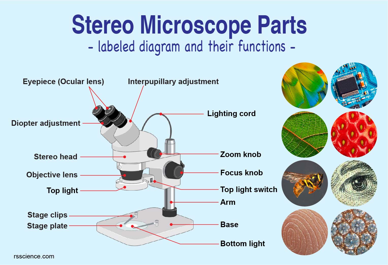
Parts of Stereo Microscope (Dissecting microscope) – labeled diagram ... Labeled part diagram of a stereo microscope Major structural parts of a stereo microscope. There are three major structural parts of a stereo microscope. The viewing Head includes the upper part of the microscope, which houses the most critical optical components, including the eyepiece, objective lens, and light source of the microscope.
Microscope diagram labeled
Microscope Labeled Pictures, Images and Stock Photos photosynthesis. Diagram of the process of photosynthesis, showing the light reactions and the Calvin cycle. photosynthesis by absorbing water, light from the sun, and carbon dioxide from the atmosphere and converting it to sugars and oxygen. Light reactions occur in the thylakoid. Calvin Cycle occurs in the stoma. Neutrophil vector illustration. Microscope Parts, Function, & Labeled Diagram - slidingmotion Microscope parts labeled diagram gives us all the information about its parts and their position in the microscope. Microscope Parts Labeled Diagram The principle of the Microscope gives you an exact reason to use it. It works on the 3 principles. Magnification Resolving Power Numerical Aperture. Parts of Microscope Head Base Arm Eyepiece Lens Cat Skeleton Anatomy with Labeled Diagram - AnatomyLearner May 29, 2021 · Cat skeleton anatomy labeled diagram. Now, I will show you all the bones from the cat skeleton with a diagram. If you find any mistakes in this cat anatomy labeled diagram, please let me know. I hope this cat skeletal system anatomy labeled diagram might help you understand and identify all the cat’s bones.
Microscope diagram labeled. Compound Microscope- Definition, Labeled Diagram, Principle, … Apr 03, 2022 · Therefore, a microscope can be understood as an instrument to observe tiny elements. The optical microscope often referred to as the light microscope, is a type of microscope that uses visible light and a system of lenses to magnify images of small subjects. There are two basic types of optical microscopes: Simple microscopes; Compound microscopes PDF Parts of a Microscope Printables - Homeschool Creations Label the parts of the microscope. You can use the word bank below to fill in the blanks or cut and paste the words at the bottom. Microscope Created by Jolanthe @ HomeschoolCreations.net. Parts of a eyepiece arm stageclips nosepiece focusing knobs illuminator stage objective lenses A Study of the Microscope and its Functions With a Labeled Diagram ... These labeled microscope diagrams and the functions of its various parts, attempt to simplify the microscope for you. However, as the saying goes, 'practice makes perfect', here is a blank compound microscope diagram and blank electron microscope diagram to label. Download the diagrams and practice labeling the different parts of these ... Interactive Bacteria Cell Model - CELLS alive Periplasmic Space: This cellular compartment is found only in those bacteria that have both an outer membrane and plasma membrane (e.g. Gram negative bacteria).In the space are enzymes and other proteins that help digest and move nutrients into the cell. Cell Wall: Composed of peptidoglycan (polysaccharides + protein), the cell wall maintains the overall shape of a …
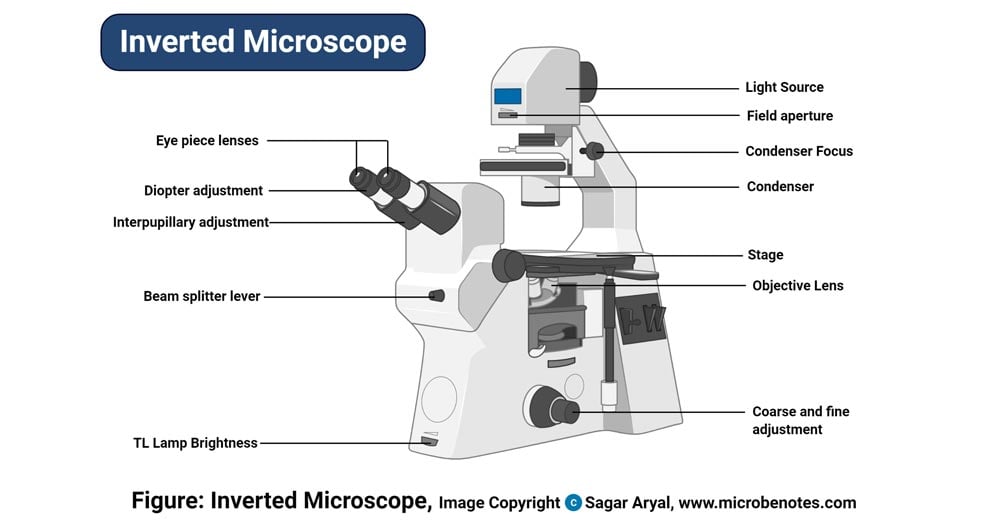
Parts of a microscope with functions and labeled diagram Sep 17, 2022 · Q. Differentiate between a condenser and an Abbe condenser. Ans. Condensers are lenses that are used to collect and focus light from the illuminator into the specimen. They are found under the stage next to the diaphragm of the microscope. They play a major role in ensuring clear sharp images are produced with a high magnification of 400X and above. Microscopy Basics | Nikon’s MicroscopyU In order to realize the full potential of the optical microscope, one must have a firm grasp of the fundamental physical principles surrounding its operation. ... Examine an animated cut-away diagram of a modern inverted microscope. ... Examining specimens labeled with molecules that absorb light and emit fluorescence. Microscope Optical Systems. compact bone diagram microscope Histology bone compact tissue connective slides human lamellae bones diagram structure anatomy cartilage ground section biology tooth bio physiology ou. Bone section structure compact histology cross cortical function tissue animal ligaments edu tendons wsu mechanical practical anatomy. Anatomy & physiology i bis 240: osteon-canaliculi Duck Anatomy - External and Internal Features with Labeled Diagram ... Jul 24, 2021 · Here, I will show you again the duck anatomy diagram as a whole so that you may summarize your contents so quickly. I tried to show you the basic anatomical features of a duck. If you need more duck labeled diagrams, please let me know. Or, you may join to anatomy learner on social media to get more updates on duck labeled diagrams.
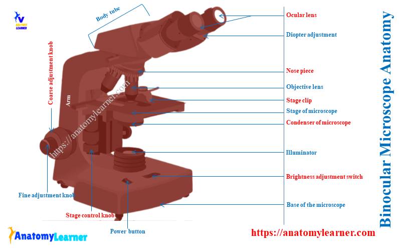
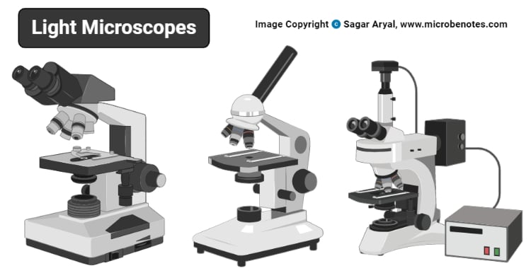
Light Microscope- Definition, Principle, Types, Parts ... Sep 07, 2022 · Parts of a microscope with functions and labeled diagram; Amazing 27 Things Under The Microscope With Diagrams; History of Microbiology and Contributors in Microbiology; 22 Types of Spectroscopy with Definition, Principle, Steps, Uses; Animal Cell- Definition, Structure, Parts, Functions, Labeled Diagram Binocular Microscope Anatomy - Parts and Functions with a Labeled Diagram Now, I will discuss the details anatomy of the light compound microscope with the labeled diagram. Why it is called binocular: because it has two ocular lenses or an eyepiece on the head that attaches to the objective lens, this ocular lens magnifies the image produced by the objective lens. Binocular microscope parts and functions Fluorescence Microscopy- Definition, Principle, Parts, Uses Apr 12, 2022 · Parts of a microscope with functions and labeled diagram; Organic waste recycling (methods, steps, significance, barriers) Limitations of Fluorescence Microscope. Fluorophores lose their ability to fluoresce as they are illuminated in a process called photobleaching. Photobleaching occurs as the fluorescent molecules accumulate chemical damage ... Label the microscope — Science Learning Hub All microscopes share features in common. In this interactive, you can label the different parts of a microscope. Use this with the Microscope parts activity to help students identify and label the main parts of a microscope and then describe their functions. Drag and drop the text labels onto the microscope diagram.
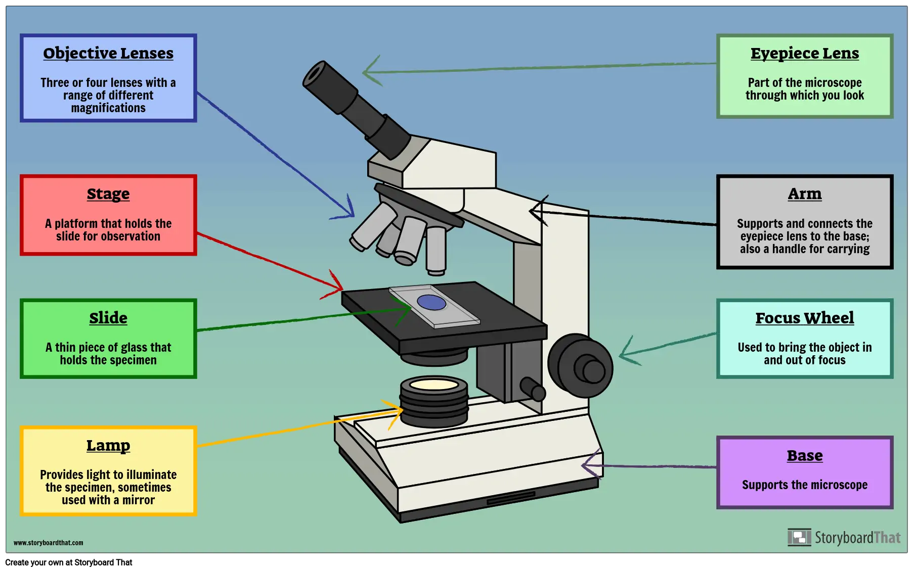
Microscope Parts and Functions With Labeled Diagram and ... Most specimens are mounted on slides, flat rectangles of thin glass. The specimen is placed on the glass and a cover slip is placed over the specimen. This allows the slide to be easily inserted or removed from the microscope. It also allows the specimen to be labeled, transported, and stored without damage.
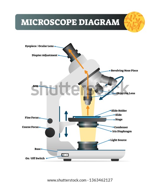
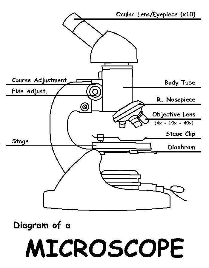
Microscope labeled diagram - SlideShare Microscope labeled diagram 1. The Microscope Image courtesy of: Microscopehelp.com Basic rules to using the microscope 1. You should always carry a microscope with two hands, one on the arm and the other under the base. 2. You should always start on the lowest power objective lens and should always leave the microscope on the low power lens ...
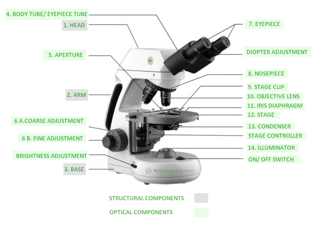
Compound Microscope Parts – Labeled Diagram and their ... Labeled diagram of a compound microscope Major structural parts of a compound microscope There are three major structural parts of a compound microscope. The head includes the upper part of the microscope, which houses the most critical optical components, and the eyepiece tube of the microscope.
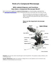
Compound Microscope Parts, Functions, and Labeled Diagram Compound Microscope Parts, Functions, and Labeled Diagram Parts of a Compound Microscope Each part of the compound microscope serves its own unique function, with each being important to the function of the scope as a whole.
Cell Nucleus – function, structure, and under a microscope [In this figure] The cell nucleus diagram and its structure. ... With the discovery of an electron microscope, we now know that the nuclear envelope is a double-layer membrane. The ball-like structure of the darker region inside the nuclei is the nucleolus, a place where ribosomes are made. ... Immunofluorescence utilizes fluorescent-labeled ...
Compound Microscope - Diagram (Parts labelled), Principle and Uses See: Labeled Diagram showing differences between compound and simple microscope parts Structural Components The three structural components include 1. Head This is the upper part of the microscope that houses the optical parts 2. Arm This part connects the head with the base and provides stability to the microscope.
Simple Microscope - Diagram (Parts labelled), Principle, Formula and Uses Parts of a Simple Microscope A simple microscope consists of Optical parts Mechanical parts Labeled Diagram of simple microscope parts Optical parts The optical parts of a simple microscope include Lens Mirror Eyepiece Lens A simple microscope uses biconvex lens to magnify the image of a specimen under focus.
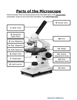
Label the microscope Diagram | Quizlet Holds the slide in place. Diaphragm. Regulates the amount of light on the specimen. Light Source. Projects light upwards through the diaphragm, the specimen, and the lenses. Arm. supports the body tube. Stage. Supports the slide being viewed.
Labelled Diagram of Compound Microscope The below mentioned article provides a labelled diagram of compound microscope. Part # 1. The Stand: The stand is made up of a heavy foot which carries a curved inclinable limb or arm bearing the body tube. The foot is generally horse shoe-shaped structure (Fig. 2) which rests on table top or any other surface on which the microscope in kept.
Microscope Types (with labeled diagrams) and Functions Simple microscope labeled diagram Simple microscope functions It is used in industrial applications like: Watchmakers to assemble watches Cloth industry to count the number of threads or fibers in a cloth Jewelers to examine the finer parts of jewelry Miniature artists to examine and build their work Also used to inspect finer details on products
Amazing Cells - University of Utah This magical microscope lets viewers jump between levels of magnification from organ systems to cells. ... After — as a check to make sure students labeled their organelles correctly. Within cells, special structures carry out particular functions. ... project to the class the Cell Signaling Steps diagram, summarizing the steps the students ...
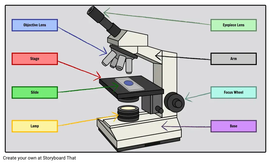
Microscope: Parts Of A Microscope With Functions And Labeled Diagram. Figure: A diagram of a microscope's components. The microscope has three basic components: the head, the base, and the arm. Head:Occasionally, the head is considered the body. It holds the optical components of the upper part of the microscope. Base:The microscope's base provides great support.
Cat Skeleton Anatomy with Labeled Diagram - AnatomyLearner May 29, 2021 · Cat skeleton anatomy labeled diagram. Now, I will show you all the bones from the cat skeleton with a diagram. If you find any mistakes in this cat anatomy labeled diagram, please let me know. I hope this cat skeletal system anatomy labeled diagram might help you understand and identify all the cat’s bones.
Microscope Parts, Function, & Labeled Diagram - slidingmotion Microscope parts labeled diagram gives us all the information about its parts and their position in the microscope. Microscope Parts Labeled Diagram The principle of the Microscope gives you an exact reason to use it. It works on the 3 principles. Magnification Resolving Power Numerical Aperture. Parts of Microscope Head Base Arm Eyepiece Lens
Microscope Labeled Pictures, Images and Stock Photos photosynthesis. Diagram of the process of photosynthesis, showing the light reactions and the Calvin cycle. photosynthesis by absorbing water, light from the sun, and carbon dioxide from the atmosphere and converting it to sugars and oxygen. Light reactions occur in the thylakoid. Calvin Cycle occurs in the stoma. Neutrophil vector illustration.



















Post a Comment for "40 microscope diagram labeled"